Non-Invasive Cardiac Investigations
An ECG is a simple and useful test which records the rhythm, rate and electrical activity of the heart. Recordings are made by connecting electrodes to your limbs and across your chest wall.
Blood pressure control his vital in preventing a number of cardiac and non-cardiac conditions. Intermittent blood pressure monitoring over a 24-hour period provides a more accurate assessment of this than single isolated recording
Continuous ECG recording during either a treadmill exercise or upright bicycle test can provide insight on problems that predominantly occur during exertion. ECG changes can identify whether there is a problem with the heart arteries and certain rhythm problems can be provoked.
Light-headedness and blackouts can sometimes be reproduced with a tilt test which can allow further assessment. Continuous blood pressure and heart rate monitoring is performed while tilted upright on a special bed under close supervision.
List of pages
-
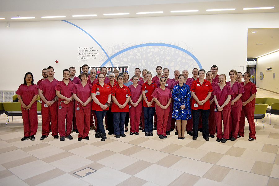
Royal Papworth Hospital's Cardiac Physiology department offers a wide range of non-invasive and invasive diagnostic procedures as well as interventional procedures for coronary disease, valve disorders, structural heart disorders, and arrhythmias.
-
Echocardiography uses ultrasound to assess the structure and function of the heart chambers and of the heart valves, providing real-time views of the heart in motion and of the blood flow through the heart.
-
Cardiac MRI is a way of creating very detailed images of the heart and surrounding structures and the high resolution of a CT scan allows assessment of the coronary arteries non-invasively. A myocardial perfusion scan uses a small amount of a radioactive substance to show blood flow to the heart muscle.